Heart

At birth or in infancy, approximately 50% of individuals with TSC (Tuberous Sclerosis Complex) have at least one tumour in their heart.
These benign (non-cancerous) tumours called cardiac rhabdomyomas do not usually cause any symptoms. Most rhabdomyomas decrease in size or disappear within the first 1-2 years of life.
The term used by health professionals for things to do with the heart is cardiac. Doctors who specialise in children’s hearts are called paediatric cardiologists.
Signs, Symptoms and
Treatments
Signs and Symptoms

Cardiac rhabdomyomas may be detected during pregnancy using standard antenatal ultrasound but may not appear until the second half of pregnancy. They may be single or multiple and are usually the first clinical clue that the baby has TSC. They are non-cancerous tumours, which means they do not metastasize, or spread throughout the body. Although it is not clear why, rhabdomyomas typically regress, or shrink, during childhood. Less commonly they can remain stable in size before they eventually become smaller and disappear.
Most individuals with TSC who have cardiac rhabdomyomas will have no symptoms.
For a smaller number of individuals, symptoms depend on the number of tumours and their size and location within the heart.
Larger tumours that are attached to the heart valves can obstruct blood flow through the heart and lead to reduced flow and symptoms depending on which valve is affected. Large tumours obstructing blood flow to the right heart and lung circulation may result in cyanosis (blue discolouration of the skin due to insufficient blood oxygenation). Large tumours obstructing blood flow to the left heart and body circulation may result in decreased perfusion to different organs and circulatory instability.
An abnormal heart rhythm, called cardiac arrhythmia can be caused when a large tumour is close to the heart’s electrical conduction system.
Large tumours that are imbedded within the heart muscles may result in poor contractility and impaired heart muscle function, or cardiomyopathy. Babies with cardiomyopathy may become quite unwell after birth and may require cardio-respiratory support, and even intravenous therapy with the aim of shrinking the size of those tumours.
A small minority of TSC patients with rhabdomyomas have a heart murmur.
Some individuals with TSC also have vascular abnormalities, which means abnormal blood vessels. These can lead to high blood pressure and very rarely other complications. High blood pressure can also be caused by kidney symptoms.
Surveillance
Fetal echocardiography (an ultrasound of the heart done during pregnancy) is recommended if rhabdomyomas have been identified on prenatal ultrasound.
Echocardiograms (a heart ultrasound) are recommended in children especially those aged less than 3 years.
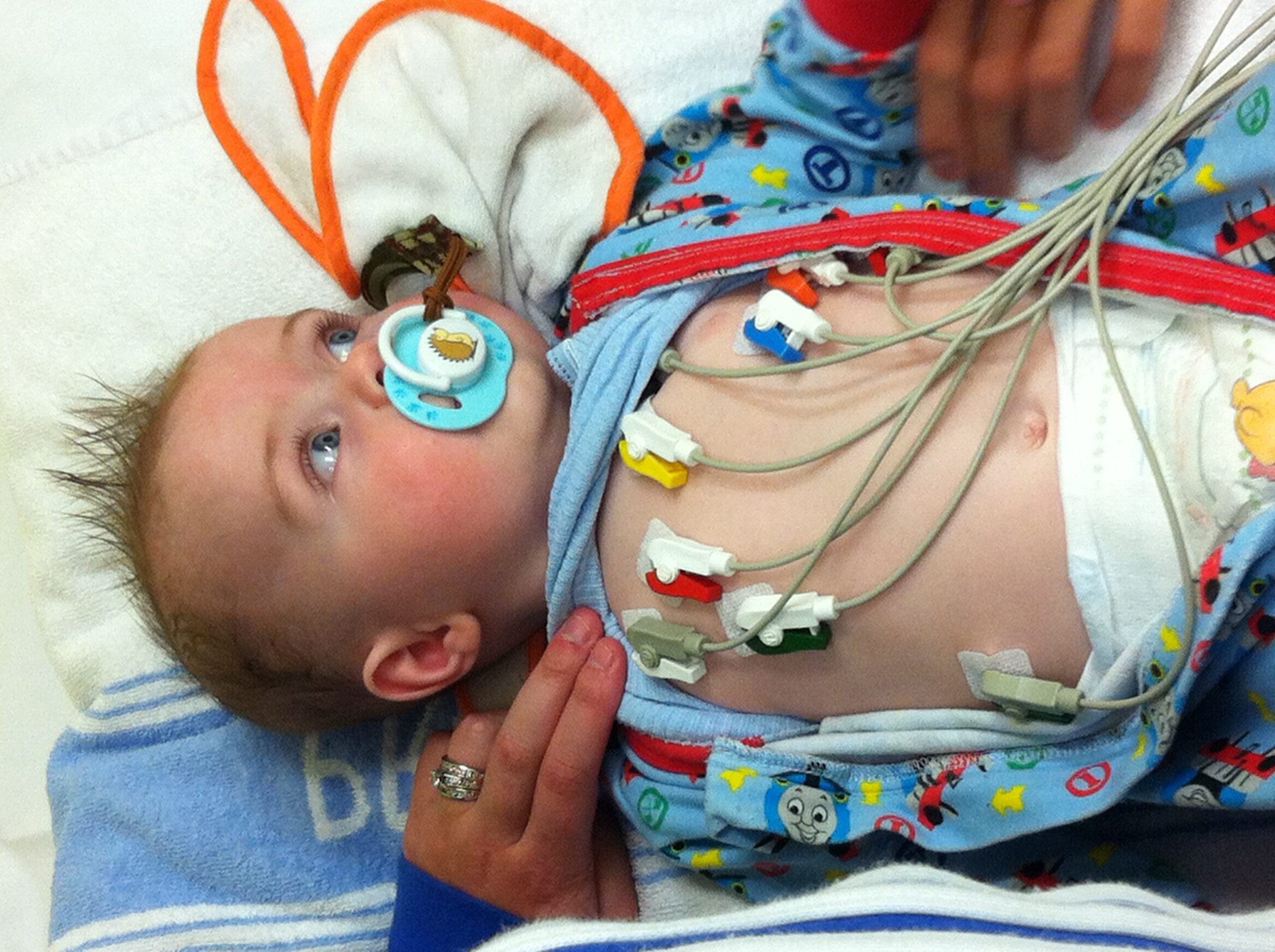
Electrocardiography (ECG) is where electrodes are placed on the skin to detect the electrical impulses of the heart to understand how well the heart conduction system is functioning. A baseline ECG should be done to diagnose any heart rhythm disturbances.
Although echocardiography is a gold standard in diagnosing rhabdomyomas in the heart, other scans such as Magnetic Resonance Imaging (MRI) and Computerised Topography (CT) may also be used to see other abnormalities in the heart.
Since heart tumours are not known to arise later in life, further imaging tests may not be necessary if initial tests did not show any tumours.
In children who have rhabdomyomas but without symptoms, echocardiograms should be done every 1-3 years and more frequently if there are symptoms of concern.
ECGs should be done every 3-5 years to monitor a person’s heart rhythm and more frequently if there are heart rhythm abnormalities from multiple or large rhabdomyomas.
If there are symptoms such as arrhythmia or unexplained loss of consciousness, then an ECG should be done to investigate the possible causes of these.
Individuals being treated with adrenocorticotropic hormone (ACTH) for infantile spasms, a form of epilepsy, should be monitored closely as there is some evidence of growth of rhabdomyomas in infants being treated with ACTH.
Smoking, high cholesterol level and high blood pressure are known risk factors for heart disease in adulthood. Patients with TSC and their family ought to try to maintain a healthy lifestyle and diet. Blood pressure should be monitored at least yearly.
Treatment
Because most rhabdomyomas regress in size, watchful monitoring may be all that is necessary in individuals who have rhabdomyomas that are not interfering with the blood flow or rhythm of the heart.
Individuals with significant arrhythmias may require treatment such as medication. There have been reports of successful use of mTOR inhibitor medicines to treat large rhabdomyomas in infancy.
Read more about mTOR inhibitor medicines.
In very rare cases where there are severe obstructive symptoms caused by large tumours, surgery to remove all or part of the tumour may be required. Because most rhabdomyomas shrink in size or even disappear, surgery is usually only done where there is a severe obstruction or a life-threatening arrhythmia.
Last updated: 16 June 2025
Reviewed by: Dr Alex Gooi, Paediatric & Fetal Cardiologist, Heart Centre for Children, the Children’s Hospital at Westmead
- Kwiatkowski D.J., Whittemore V.H. & Thiele E.A. (2010) Tuberous Sclerosis Complex: Genes, Clinical Features, and Therapeutics. Weinheim: Wiley-Blackwell
- Heart Manifestations in TSC, TSC Alliance, viewed 30th March 2012
Parts of this web page have been adapted with permission from copyrighted content developed by the TSC Alliance, https://.tscalliance.org/www.tsalliance.org.
